Bilateral Basal Ganglia Calcification Causes Radiology

Basal Ganglia Calcification Radiology Reference Article
Basal Ganglia Calcification Radiology Reference Article
Cureus Bilateral Basal Ganglia Calcification Fahr S Disease

Basal Ganglia Calcification Radiology Reference Article
Basal Ganglia Calcification Radiology Reference Article
Extensive Basal Ganglia Calcification Radiology Case

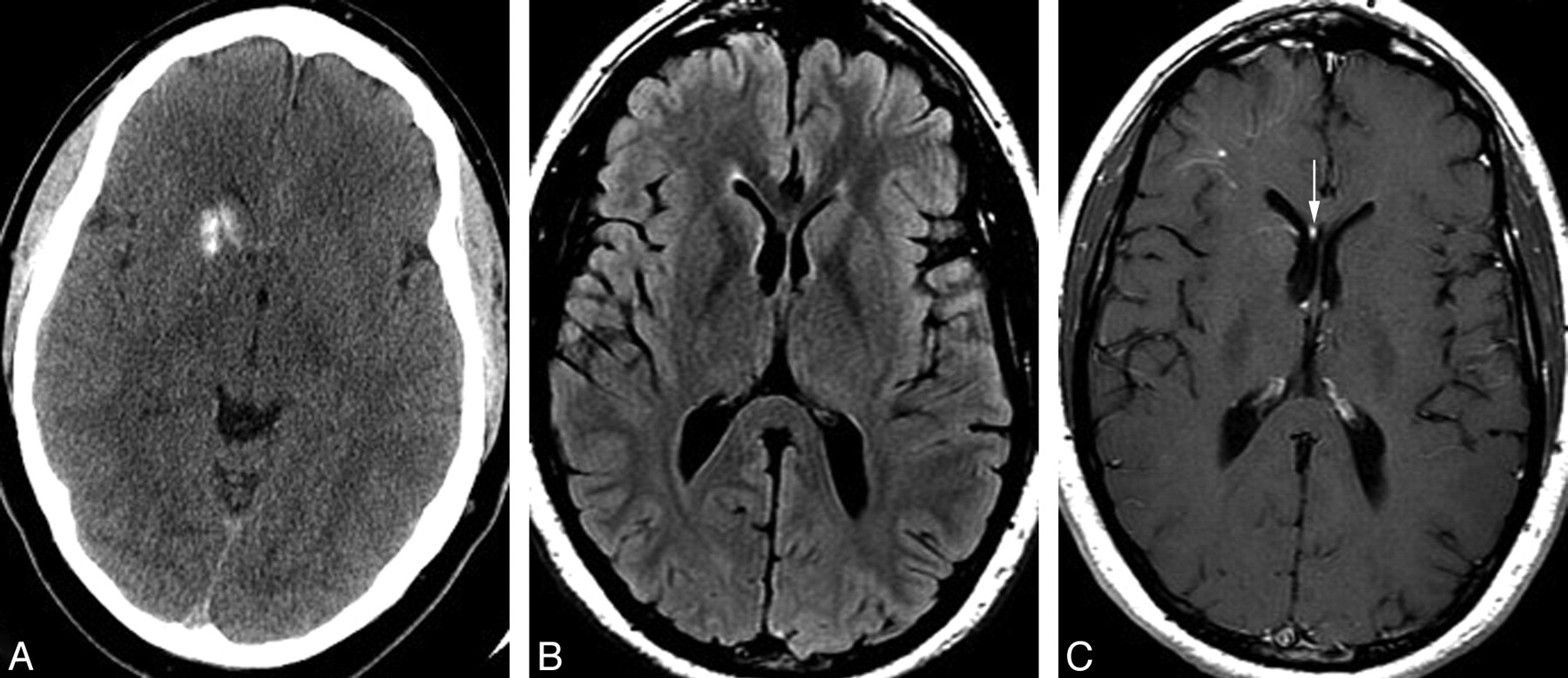
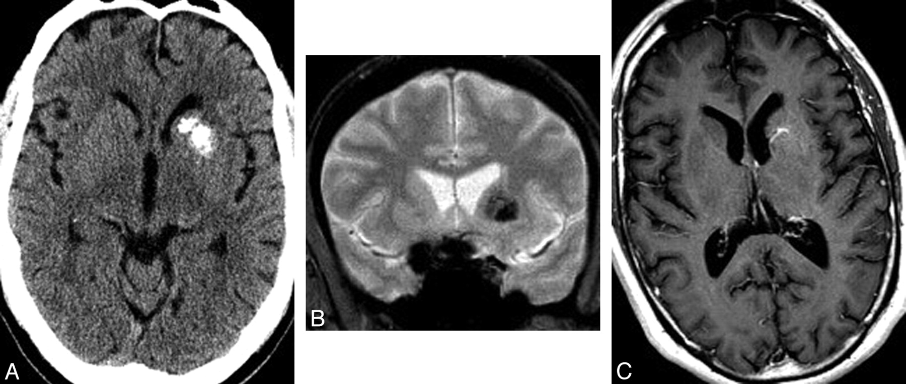
The axial t2 sequences showed bilateral hypo intensity with hyper intensity in t1w images at the medial and lateral segments of the globus pallidus separated by a thin hyperintense band on t2 fig 2.
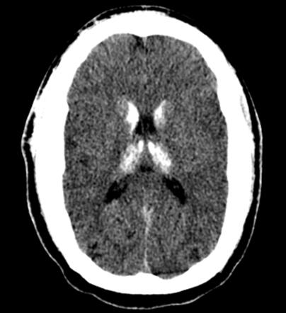
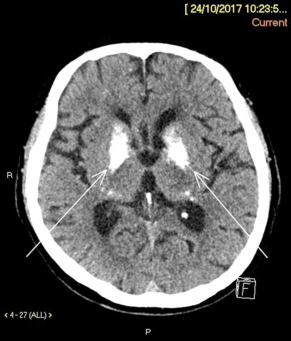
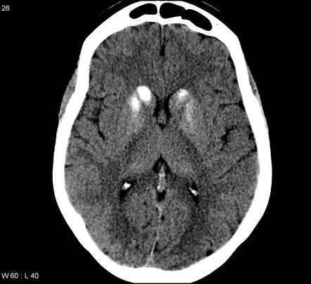
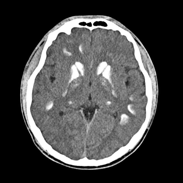
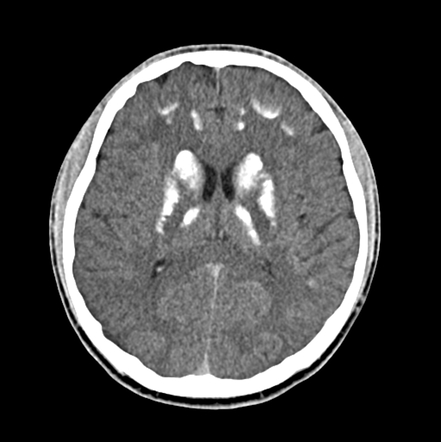
Bilateral basal ganglia calcification causes radiology. However there are many causes of calcification. Common globus pallidus most commonly affected. Bilateral basal ganglia calcification was first recognized as a histologic entity in 1855 and described independently in that year by virchow 1 and bamberger 2. Mri the mri appearance varies depending on the degree of calcification and the stage of the disease.
Bilateral thalamic lesions are usually seen in combination with basal ganglia white matter and sometimes cortical lesions. Underestimated drawbacks of combined simple dilation and thrombolytics for restoration of thrombosed brescia cimino. Causes basal ganglia calcification sometimes happens when you age but many times comes from genes passed to you by your parents. Vascular deep cerebral vein thrombo.
You only need one faulty gene from one parent to get the illness. Radiologists may detect bilateral abnormalities of the basal ganglia and thalamus in different acute and chronic clinical situations and although magnetic resonance mr imaging is the modality of choice for evaluation the correct diagnosis can be made only by taking all relevant clinical and laboratory information into account. 1971 ellie et al. 1989 manyam 2005 such as bilateral calcification of the basal ganglia visualized on neuroimaging progressive neurologic dysfunction absence of biochemical abnormalities infectious toxic or traumatic cause and family history consistent with autosomal.
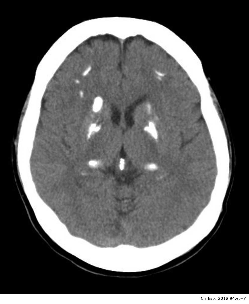
This latter characteristic mr brain pattern can have an essential diagnostic role in the early manifestation of fucosidosis. Certain diagnostic criteria are derived from moskowitz et al. Bilateral calcification of the basal ganglia on neuroimaging or other brain regions although in isolated cases patients from families with fibgc may not present such findings. Associated with radiation therapy and chemotherapy.
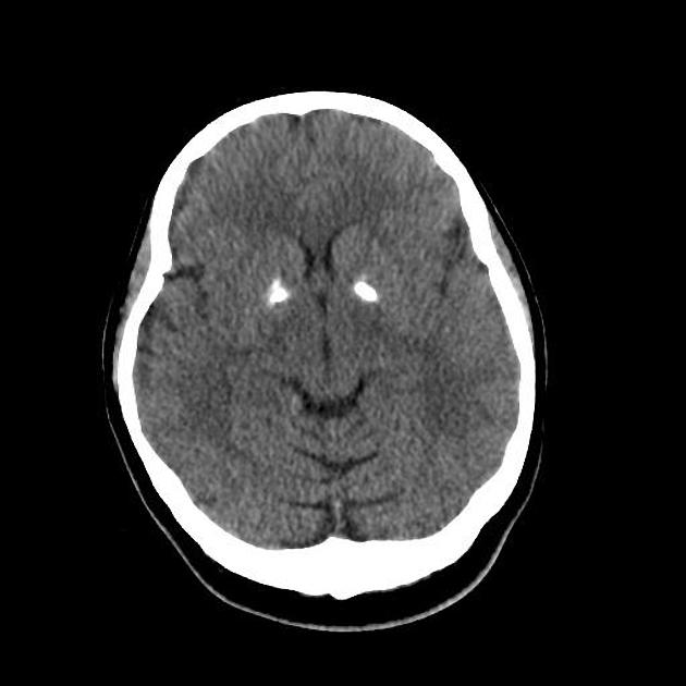
The basal ganglia and thalami are paired deep grey matter structures with extensive metabolic activity that renders them susceptible to injury by various diseases. Symmetrical bilateral involvement of the thalami has a broad differential diagnosis.

Basal Ganglia Calcification In Idiopathic Hypoparathyroidism
Extensive Basal Ganglia Calcification Radiology Case
Basal Ganglia Calcification Down Syndrome Radiology Case

Basal Ganglia Calcification In A Case Of Pkan Semantic Scholar

Dr Balaji Anvekar Frcr Intracranial Calcifications
Unilateral Calcification Of The Caudate And Putamen Association

Quick Radiology Causes For Basal Ganglia Calcification Youtube
A Unique Presentation Of Fahr Syndrome Secondary To Chronic

Bilateral Basal Ganglia Calcification And Recurrent Generalized
Brain Calcifications And Primary Hyperparathyroidism Cirugia

Full Text Fahr 39 S Disease Current Perspectives Odrr

Mri Brain Showing Bilateral Basal Ganglia Calcification
Unilateral Calcification Of The Caudate And Putamen Association
