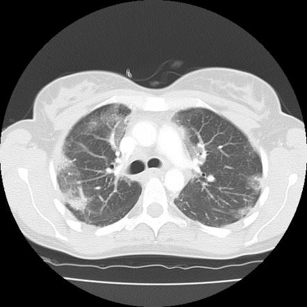
Bilateral Basal Ground Glass Opacities

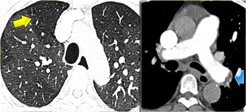
Ground glass opacification opacity ggo is a descriptive term referring to an area of increased attenuation in the lung on computed tomography ct with preserved bronchial and vascular markings.
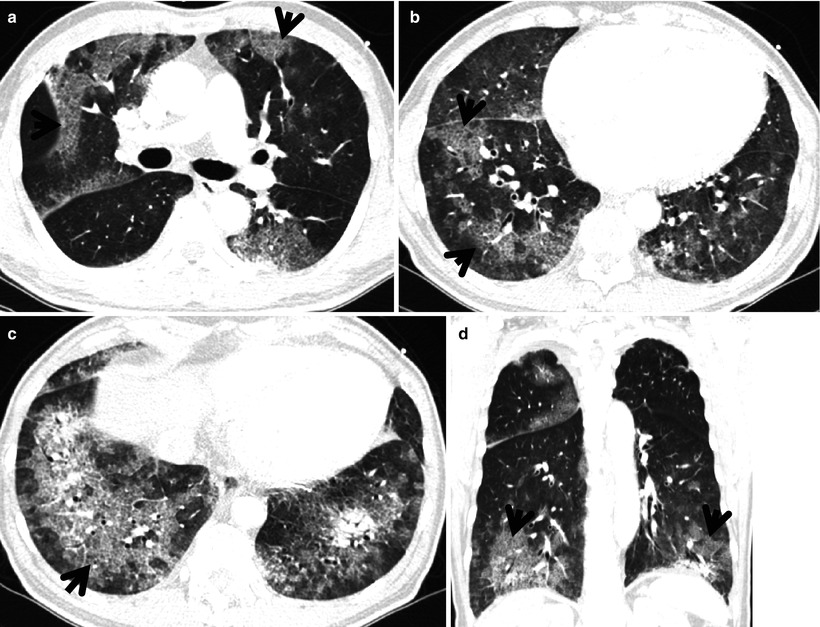
Bilateral basal ground glass opacities. Bilateral peripheralbasal dependent ground glass opacities ggos are the hallmark especially early in the disease. Ground glass opacity ggo is the descriptive term used to refer to this hazy area. The fact that both the airspaces and interstitial tissues are often involved should have little importance when evaluating radiographs or high resolution ct hrct images. Some patients can have a rounded appearance of opacities.
Keep reading to find out more about ground glass opacities and some specific treatment options. It is a non specific sign with a wide etiology including infection chronic interstitial disease and acute alveolar disease. In radiology ground glass opacity is a nonspecific finding on radiographs and computed tomography scans. Bilateral abnormalities filling of alveoli enlarged heart rapid response to diuretics ground glass opacity due to filling of alveoli with fluid gravitational distribution of the alveolar fluid.
Ground glass opacification has therefore been categorized as nonspecific by many radiologists. It usually has preserved vascular and bronchial markings as well and may well be the result of an acute alveolar disease. Chest ct in covid 19 pneumonia demonstrates bilateral peripheral and basal predominant ground glass opacities ggos and or consolidation in nearly 85 of patients with superimposed irregular lines and interfaces. The typical hrct features of aip are bilateral multifocal or diffuse areas of ground glass opacity and consolidation usually without pleural effusion efig.
Consolidations can be seen in the later stages but rare to see them without ggos later stages can show crazy paving appearance reverse halo atoll sign. Acute bilateral airspace opacification is a subset of the larger differential diagnosis for airspace opacification. It consists of a hazy opacity that does not obscure the underlying bronchial structures or pulmonary vessels and that indicates a partial filling of air spaces in the lungs by exudate or transudate as well as interstitial thickening or partial collapse of lung alveoli. The imaging findings peak at 9 13 days post infection 7 8 figure 1.
The role of the radiologist is evolving and is becoming more significant in.


