Bilateral Diffuse Ground Glass Opacities Ct

Ground Glass Opacity Wikipedia

Ground Glass Opacification Radiology Reference Article

Ct Scan Of Chest Showing Bilateral Diffuse Ground Glass Opacity Of

Ct Scan Of Thorax Of A Patient Showing Diffuse Ground Glass

Ground Glass Opacity Wikipedia

An Unusual Cause Of Patchy Ground Glass Opacity Thorax

The endoscopic findings did not reveal any endobronchial lesions.
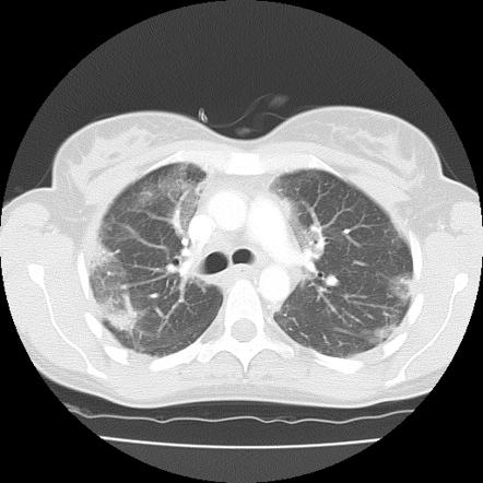
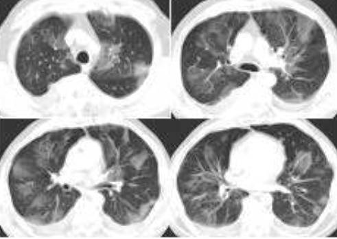
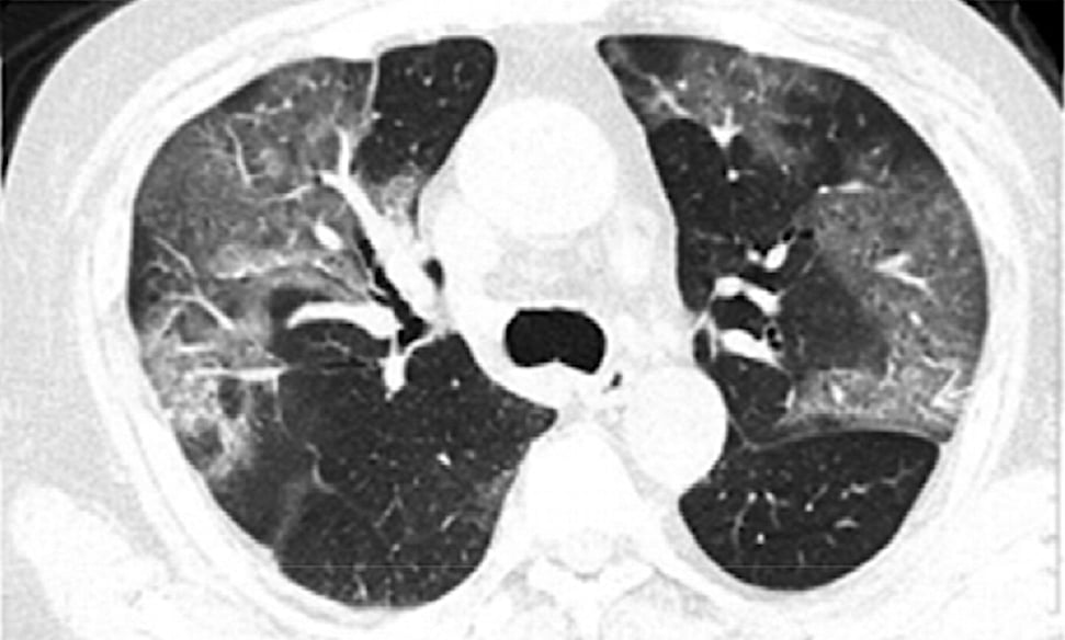
Bilateral diffuse ground glass opacities ct. Carinii and cmv pneumonias affect similar populations often have similar imaging characteristics and often cannot be distinguished on the basis of imaging. The imaging findings peak at 9 13 days post infection 7 8 figure 1. Covid 19 pneumonia manifests with chest ct imaging abnormalities even in asymptomatic patients with rapid evolution from focal unilateral to diffuse bilateral ground glass opacities that progressed to or co existed with consolidations within 1 3 weeks. High resolution ct image through inferior hilum shows isolated diffuse ground glass opacity widely spread across both lungs.
82 83 ground glass opacities reflect underlying alveolitis. It is a non specific sign with a wide etiology including infection chronic interstitial disease and acute alveolar disease. Flexible bronchoscopy and transbronchial biopsy of the lung parenchyma and bronchoalveolar lavage bal were performed. In acute hp chest imaging typically reveals diffuse ground glass opacities although a fine micronodular pattern may also be observed fig.
Pure ground glass opacities do not have solid components but you can also develop part solid ggos that are a combination of both ggo and a solid component. Chest ct in covid 19 pneumonia demonstrates bilateral peripheral and basal predominant ground glass opacities ggos and or consolidation in nearly 85 of patients with superimposed irregular lines and interfaces. It consists of a hazy opacity that does not obscure the underlying bronchial structures or pulmonary vessels and that indicates a partial filling of air spaces in the lungs by exudate or transudate as well as interstitial thickening or partial collapse of lung alveoli. 84 paralleling the clinical response radiographic abnormalities in acute hp resolve over days to weeks if further exposure is avoided efig.
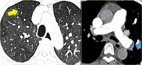
Due to the availability of better technology and widespread use of computed tomography ct it is quite common now to encounter pulmonary nodules with ggo in routine clinical practice. Ground glass opacification opacity ggo is a descriptive term referring to an area of increased attenuation in the lung on computed tomography ct with preserved bronchial and vascular markings. Although they can be seen in any stage of hp ground glass opacities are the predominant finding in acute hp. In radiology ground glass opacity ggo is a nonspecific finding on radiographs and computed tomography ct scans.
With bilateral ground glass opacities ggo resembling in terstitial pneumonia on chest computed tomography ct. Thus based on the autopsy we posthu mously diagnosed the patient with pte.
Ground Glass Opacification Radiology Reference Article

Chest Ct Scan Showing Diffuse Patchy Ground Glass Opacities Mainly

Ground Glass Opacity An Overview Sciencedirect Topics

Differential Diagnosis And Management Of Focal Ground Glass

Chest Radiology
Ct Scan Of Chest Ground Glass Opacification Youtube

Figure 1 From Clear Vision Through The Haze A Practical Approach

Chest Ct Scan Shows Bilateral Diffuse Ground Glass Opacities With

Covid 19 Round And Oval Areas Of Ground Glass Opacity Pulmonology

Predominant Diffuse Ground Glass Opacity In Both Lung Fields A
Southwest Journal Of Pulmonary Critical Care Imaging Ground
Chest Ct Findings Of Patients Infected With Novel Coronavirus 2019

Differential Diagnosis And Management Of Focal Ground Glass